CT scans are important diagnostic tools in medicine. They are generally performed in radiology departments or departments for diagnostic imaging in hospitals. They are extremely useful to get detailed information that surpasses the simple findings an x-ray can provide.
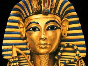
Recently CT scanning has been used as a tool by archeologists to examine a patient that has passed away 3,300 years ago. Tutankhamun, the Egyptian king, died very young. After an x-ray examination in 1968 which seemed to detect bone fragments in the boy king’s skull, it was speculated that he had been a victim of foul play. Dr. Ashraf Selim, a radiologist at Cairo University and leader of the CT examination of King Tut, did not find any evidence of this. During the discovery of the mummy by the Englishman Howard Carter in 1922 Carter and his cronies were quite rough, when they tried to remove the pharaoh’s golden mask, and as a result some bone fractured, which also matched a defect within the first cervical vertebra. This being an injury long time after death excluded foul play. What was obvious in the CT finding was a fracture to the femoral bone, which occurred before the death of the young king. While researchers cannot assess how this injury happened, the findings suggest that the injury was likely an open wound that became infected and led to the untimely death of the king (no antibiotics there at that time).
It is rare that archeologists will draw on CT scans to uncover a mysterious death, but CT scans are not only tools for specialists like orthopedic surgeons or neurologists. They can be a helpful tool to assist in other areas of medicine such as forensic medicine to find valuable insights.
Reference: The Medical Post, January 16,2007, page 16
Last edited December 5, 2012


