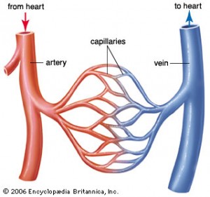
Researchers at the Duke University Medical Center have developed a new non-invasive method of visualizing blood flow through blood vessels of patients. It is a modification of the well-known MRI scan technology where a magnetic field realigns the center of hydrogen atoms (protons) during the time of the examination and the differences of the tissue and fluid qualities are reflected in the images created by this technology. This new application depicting moving blood in blood vessels is called “global coherent free procession” (GCFP).
The principle is that the investigator can focus on an area of a blood vessel upstream of the area to be examined and tag a portion of the blood flowing through with an energy pulse. As the blood continues to flow through the area of interest, the protons give off the energy again without any changes to the body fluids or the blood cells and the MRI scanner picks up the images of the blood flow through the blood vessel.
The advantage of this technique is that it is done without any catheters (it is non-invasive), there is no need for any contrast material to be injected and there are no X-rays needed. At the present time this is the only diagnostic technology available for examining a patient’s blood flow through the heart and its vessels in real time, which is very valuable for physicians (cardiologists).
Here is a link to the Duke University publication
Based on an article in the April 13, 2004 issue of The Medical Post , Vol.40, No. 15, p.5.
PS. When checked on Nov. 5, 2012 this procedure has not been widely accepted in medical circles. It seems to be still more of a research tool.
Last edited December 8, 2012


