Nov
07
2020

Removal of Senescent Cells Can Extend Life

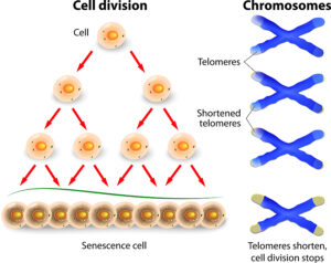
Several animal and human studies by the Mayo Clinic showed that removal of senescent cells can extend life. Researchers Xu et al. showed in 2018 that senescent cells weaken the body. Senescent cells are damaged cells that are still living. They can cause the release of inflammatory cytokines. The researchers showed in mouse experiments that intermittent senolytics increased life expectancy by 36%. Senolytics are drugs that dissolve senescent cells; the senolytic cocktail consisted of dasatinib plus quercetin.

In these mouse experiments their risk of dying was reduced by 65% compared to control mice that did not take senolytics.

Senescent cells causing premature aging

In the past 5 years research on aging and on chronic diseases made a lot of progress. Researchers realized that the accumulation of senescent cells is what causes both. All this happens because the process of apoptosis, the removal of dead cells, is impaired in the aging person. It appears that in older age there is a problem with dying cells and their removal. Instead they linger on and start producing cytokines, which cause inflammation. This can damage other cells and lead to organ failures. All this explains why older people often get chronic diseases and do not reach their normal lifespan. The accumulation of senescent cells also blocks regenerative factors that improve one’s health.

Senolytics

Dasatinib is a kinase inhibitor that was developed to treat acute myelogenous leukemia in adults and children. Researchers did animal experiments with a combination of dasatinib and quercetin for several years. They also have started smaller pilot clinical trials in humans. It appears that the human findings are very similar to the animal findings. But more research is needed to answer questions about side-effects and effects of removal of senescent cells.

Details about animal experiments with senolytics

The Mayo Clinic research showed that old mice treated with senolytics (dasatinib and quercetin) live 36% longer than controls that did not receive senolytics. Another part of this series of experiments showed that senescent cells are indeed what kills prematurely. They took senescent cells from old mice and transplanted them into young mice. Soon the young animals showed deterioration health wise and they died prematurely. Another control group were older mice that received senescent cells from old mice. They too died prematurely. Treatment with senolytics (dasatinib and quercetin) improved physical functioning and also survival.

Details about human trials regarding senolytics

For three days 11 participants received senolytics (dasatinib and quercetin). The effect of the drugs was evident for 11 days. The subjects took 100 mg of dasatinib daily and 500 mg of quercetin twice per day for 3 consecutive days. This dose was repeated twice more on a weekly basis for a total of 3 weeks. These patients had idiopathic pulmonary fibrosis. This is an incurable disease where senescent cells accumulate. These patients showed significantly improved gait speed, walking endurance, chair rise test performance and scores of other physical performances. All this occurred on day 5 after the initial dose of senolytics.

Alternative senolytics, so removal of senescent cells can extend life

Dasatinib as a senolytic has significant side effects.

For this reason, researchers looked for alternatives. Theaflavins, isolated from black tea fits this bill. It is non-toxic, but it is also effective as a senolytic. Researchers from Life Extension have developed a senolytic product containing theaflavins and quercetin. Instead of regular quercetin they included quercetin phytosome, which has 50-times more potent bioavailability. One capsule contains 74 mg of quercetin phytosome (the equivalent of 1250 mg of regular quercetin) and 275 mg of theaflavins.

Discussion

Future research needs to show whether or not the Life Extension senolytic indeed does what it promises. It claims that only one capsule per week stimulates apoptosis, reduces cytokines and increases longevity. I would like to see a clinical study that examines all these parameters. One measure of longevity is to determine the length of leukocyte telomeres. All the other laboratory tests are readily available. Research in this field will certainly continue and scientists will likely develop other senolytics.

Removal of Senescent Cells Can Extend Life

Removal of Senescent Cells Can Extend Life

Conclusion

The accumulation of senescent cells causes both aging and chronic diseases. Research showed that in older age the process of apoptosis, the removal of dying cells, is incomplete. As a result dying cells accumulate. They produce inflammatory cytokines, can damage other healthy cells and lead to chronic organ failure. In addition, cancer cells can develop and the patients can die prematurely. Senolytics are substances that clear out senescent cells. In mouse experiments they have already led to improved survival and health. Clinicians performed a clinical trial on patients with idiopathic pulmonary fibrosis, which is an incurable disease where senescent cells accumulate. They showed significantly improved gait speed, walking endurance, chair rise test performance and scores of other physical performances. One pill once per week with dasatinib and quercetin can achieve this. More research in this area can clarify why senolytics work and what the side effects are.
Oct
31
2020

Blood Type Has Some Bearing on the Severity of Covid-19 Coronavirus

Two independent research publications concluded that blood type has some bearing on the severity of Covid-19 coronavirus infections. One was published in Denmark, the other one in Canada.

In the US the 4 common blood types occur with this frequency: group O: 45% (O positive 38%, O negative 7%); group A:  40% (A positive 34%, A negative 6%); group B: 11% (B positive 9%, B negative 2%) and group: AB 4% (AB positive 3%, AB negative 1%). Positive and negative stands for the Rh group (the rhesus factor, which is another type of blood group).

Two separate publications

Denmark study

Briefly, the Denmark study showed that when positive and negative tests for the SARS-CoV-2 virus were checked in relationship to blood groups, blood group O had 13% less coronavirus infections, group A had 9% more infections, group B had 6% more infections and group AB had 15% more infections than negative controls. This means that blood group O is relatively protected from the SARS-CoV-2 virus. The investigators were fast to add that this does not give people with a group O blood type a licence to go to the pub and celebrate.

Canadian study

The Canadian study looked at 125 critically ill people with positive SARS-CoV-2 virus tests. Of these 95 had ABO blood types available. All these patients were admitted to the ICU. Here are the significant findings: 32% of blood group A required intubation versus 84% of AB patients, 35% of O group patients and 61% of B patients required intubation. 12% of A patients and 32% of AB patients, but only 5% of blood group O patients and 9% of B patients required kidney support (continuous renal replacement therapy). In addition, group O and group B patients required a median ICU stay of only 9 days. In contrast, group A and AB had to stay in the ICU for 13.5 days.

Gene study to determine susceptibility for severe Covid-19 disease

In a European genetic study from Italy and Spain 835 patients and 1255 control participants had genetic studies done. It turned out that the genetic loci that determined the severity of Covid-19 followed the blood groups. Blood group A patients had a 45% higher risk of developing severe Covid-19 disease, while group O patients had a 35% lesser risk compared to other blood groups of developing severe Covid-19.

Discussion

Dr. Mypinder Sekhon, an intensive care physician at Vancouver General Hospital stated that people with a blood group O make less of a key clotting factor, which makes them less prone to clotting problems in the blood. Clotting is a major driver in complications of Covid-19. Other possible explanations are the blood group antigens and how they interact with antibodies from the infection with Covid-19 coronavirus. Finally, it could be related to the genes of the blood groups and how they interact with receptors of the immune system. It is interesting to also note that there are genetically different risks that go along the line of the blood groups with group A having much higher risk than group O to develop severe Covid-19 disease.

Blood Type Has Some Bearing on the Severity of Covid-19 Coronavirus

Blood Type Has Some Bearing on the Severity of Covid-19 Coronavirus

Conclusion

Both research in Denmark and in Canada confirmed that blood group O had less coronavirus infections. In the Denmark study there were 13% less severe Covid-19 cases in people with the blood group O than in negative controls. In the Canadian study of 95 patients with severe Covid-19 physicians had to admit them into the ICU. They noted that 84% of blood group AB patients in the ICU required intubation. In contrast, only 35% of O group patients required intubation. With regard to kidney support, 32% of AB patients, but only 5% of O patients required this during their ICU stay.

More research required to understand these findings

The researchers added that it is not clear why there are such differences among patients with different blood groups. They mentioned that more research is necessary. This will reveal why group O has a milder course. It will also show why group O patients require less intubation and shorter ICU stays. Separate genetic studies showed that severe Covid-19 disease develops with blood group A patients (45% higher risk). In contrast group O blood group patients have milder Covid-19 disease (35% less risk). It is with these investigations that we can now understand some of the peculiarities regarding the Covid-19 disease.  It explains why some people develop severe Covid-19 disease while others develop only mild symptoms.

Sep
05
2020

How to Manage Clot Formation with Covid-19

A publication in the Canadian Medical Association Journal describes how to manage clot formation with Covid-19. A significant amount of cases among Covid-19 patients come down with clotting problems. This means that an infection with SARS-CoV-2 (or Covid-19 coronavirus) may initially present with a fever and cough. But a few days later it can suddenly turn into a dangerous disease with severe clots, multiple organ failures and death.

Clot occurrence with Covid-19

It is important to realize that most patients with SARS-CoV-2 do not need hospitalization. But physicians admit 10 to 15% of patients to the hospital. Of these 20% end up with treatment in the Intensive Care Unit (ICU). Of all the hospitalized patients between 5% and 30% develop some form of thrombotic event. Notably, complications of clot formation can be a stroke, a heart attack, a pulmonary embolism or a deep vein thrombosis in the leg. In a recent study from the US 400 random hospitalized patients with Covid-19 144 patients were admitted to the ICU. 4.8% had radiologically confirmed deep vein thrombosis. Overall there were 9.5% with thrombotic events that developed during the hospital stay.

How does a coagulopathy develop with Covid-19?

Truly, SARS-CoV-2 enters the body cells through an interaction of its viral spike protein with the angiotensin-converting enzyme 2 (ACE2) receptor. To explain, numerous organs and tissues express this receptor. This includes lung alveolar type 2 epithelial cells, endothelium, the brain, heart and kidneys. To emphasize, ACE2 leads to angiotensin II degradation. With the SARS-CoV-2 stimulation of the ACE2 receptor there may be an accumulation of angiotensin II, which causes a procoagulant state. Injury of the endothelium explains inflammation in the lining of the blood vessels in multiple organs. Commonly affected organs are lungs, heart, kidneys and intestines. The inflammatory reaction is what can lead to clot formation. When part of an organ has died off because of mini clots that destroyed part of the organ, this process can eventually lead to organ failure. Lung failure, heart failure and kidney failure can develop in these sick patients.

Adequate vitamin D blood levels are important for the immune system

By all means, vitamin D is very important for the integrity of the immune system. With vitamin D blood levels below 15 to 20 ng/mL (37.5–50 nmol/L) the immune system is paralyzed, and any viral or bacterial infection tends to overwhelm the body. Of course, this is the reason why the mortality due to Covid-19 coronavirus is highest in patients with these low vitamin D blood levels. People with secondary illnesses (diabetes, arthritis, autoimmune diseases, cancer) and patients above the age of 60 have the lowest vitamin D blood levels and have the highest mortality rates. This publication describes this in more detail.

Best vitamin D blood level is in the upper normal range (50-80 ng/mL)

Above a vitamin D blood level of 30 ng/mL (=75 nmol/L) a patient’s immune system is functioning normally. However, the immune system is strongest at a vitamin D blood level of 50–80 ng/mL (125–200 nmol/L), which is the upper range of the normal level for vitamin D in the blood.

Keep in mind that vitamin D toxicity occurs only above 150 ng/mL (375 nmol/L).

Specific effects of vitamin D on Covid-19

There are three major effects that vitamin D has.

  1. A strengthening of the epithelial barrier not allowing the coronavirus to penetrate into the lung tissue as easily.
  2. Release of defensins and cathelicidin, two crucial antiviral polypeptides that eradicate any virus in the system.
  3. Interruption of the “cytokine storm”, an overwhelming inflammation which is responsible for viral pneumonia to develop. Without the cytokine storm there is no damage to the lungs and people do not need treatment in the ICU. This is particularly important for people above the age of 60 and for people with pre-existing diseases.

In like manner, with the stabilizing effect of vitamin D regarding the immune function more severe forms of Covid-19 can turn into less severe forms with a better outcome.

Treatment of patients with Covid-19 who have clotting problems

Patients need to be assessed with respect to their risk of developing clots. This publication describes that high risk patients have elevated D-dimer levels. When blood clots dissolve the body produces D-dimer, a protein fragment. Normally the D-dimer test is negative in a person that does not produce clots. But in sick patients with Covid-19 who form clots this blood test typically shows D-dimer >2500 ng/mL. In addition the tests show high platelet counts (more than 450 × 109/L), C-reactive protein (CRP) >100 mg/L and an erythrocyte sedimentation rate (ESR) >40 mm/h.

Indeed, with this constellation of blood tests in a severe Covid-19 case in the ICU setting, the physician uses heparin intravenously or subcutaneously to counter clot formation. However, this needs to be balanced against the risk of causing severe internal bleeding.

Separate from the anticoagulant effect, heparin seems to also suppress inflammatory cytokine levels. In addition, heparin suppresses neutrophil chemotaxis and migration. Physicians rescued many patients from death using heparin therapy.

Risk versus benefit clinical trials of heparin therapy are required

At this point there are only retrospective clinical trials available to describe risk versus benefit of heparin therapy. Some show no difference, others do. There are two international clinical trials on their way to shed more light on this situation. Until the results of these clinical trials are available, physicians need to treat patients to the best of their knowledge.

How to Manage Clot Formation with Covid-19

How to Manage Clot Formation with Covid-19

Conclusion

Clot formation in sick Covid-19 patients is responsible for many deaths in Covid-19 patients. The SARS-CoV-2 (or Covid-19 coronavirus) causes a cytokine storm with injury to the lining of the arteries. This can affect multiple vital organs and the condition may lead to organ failure. This activates the clotting system and causes clots all over the body. When this process occurs, patients get very sick and the death rate climbs. Physicians were able to rescue some patients with heparin therapy. Two international clinical trials are on the way. Hopefully  these trials answer questions about this newer treatment method. The downside of heparin therapy is the complication of massive bleeding, which causes deaths as well. When it comes to Covid-19, don’t rely on curative medicine. Strengthen your immune system by preventative therapy like vitamin D3 that can interrupt the cytokine storm.

And even with a “well-prepared” immune system it is extremely important to follow all the guidelines of distancing, disinfecting and wearing face masks. We need all the help we can get!

Aug
01
2020

Eating Fish Protects the Brain from Air Pollution

Research on white women aged 70 years or older found that eating fish protects the brain from air pollution. Dr. Ka He from the Columbia University of New York published the new study on July 15, 2020 in the online issue of Neurology®, the medical journal of the American Academy of Neurology.

1,315 women who did not have dementia at the start of the study enrolled in it. Researchers measured the air pollution of the areas where the women lived. They also measured their omega-3 fatty acid blood levels. They found that women from polluted areas who ate the lowest amounts of fish and had the lowest amounts of omega-3 fatty acids in their blood, had the highest amount of brain shrinkage.

Methods of determining brain shrinkage, omega-3 fatty acids and pollution

The amount of brain shrinkage was determined with MRI scans of the brain. The amount of white brain matter was measured, particularly the size of the memory-sensitive hippocampus. Blood omega-3 fatty acid content was determined in red blood cells. Pollution was determined by the fine particulate matter in air pollution at the address where the patient lived. Every woman in the study received a diet questionnaire regarding fish consumption. From this information the researchers determined the average fish consumption per week. This included broiled and baked fish, non-fried shellfish, canned tuna, tuna casserole and tuna salad. Deep fried fish was not part of the list, because other studies showed that deep-frying damages omega-3 fatty acids.

More details regarding the study

The researchers adjusted the study according to age, smoking status, and other factors that could affect brain shrinkage. Women with the highest intake of omega-3 fatty acids had the highest volume of white matter in their brains as MRI scans showed. Specifically, the researchers noted the following findings:

  • Women with the highest omega-3 fatty acid level had 410 cubic cm white matter
  • Women with the lowest omega-3 fatty acid level had 403 cubic cm white matter
  • Each quartile increase in air pollution caused shrinkage of white matter by 11.52 cubic cm in those women with low omega-3 fatty acid consumption
  • In women with higher omega-3 fatty acid levels each quartile increase in air pollution caused shrinkage of white matter by only 0.12 cubic cm
  • Women with the highest omega-3 fatty acid intake had the highest volume of the hippocampus

Comments by the lead author of the study

Dr. Ka He stated: “Our findings suggest that higher levels of omega-3 fatty acids in the blood from fish consumption may preserve brain volume as women age and possibly protect against the potential toxic effects of air pollution.” But Dr. Ka He cautioned: “It’s important to note that our study only found an association between brain volume and eating fish. It does not prove that eating fish preserves brain volume. And since separate studies have found some species of fish may contain environmental toxins, it’s important to talk to a doctor about what types of fish to eat before adding more fish to your diet.”

Limitations of study

The study was involving older white women. This means that the results cannot be generalized to Afro Americans, Hispanics or Asians. The researchers examined exposure to pollution only later in life, not in early life or midlife. Hopefully future studies will examine what happens with lifelong exposure to pollution.

How to limit mercury exposure when eating fish

Omega-3 fatty acids are abundantly present in fish. It has plaque-reducing properties and also reduces the risk for abnormal heart beats. Overall this means less cardiovascular disease. The American Heart Association recommends a 3.5 oz. serving of fatty fish (salmon, mackerel, lake trout, herring or sardines) twice per week.

Mercury and other pollutants

Pollution of the air, soil and rivers is causing accumulation of mercury and other heavy metals in ocean water.

This affects fish that live in the ocean. There is a pecking order of predators with the larger fish feeding on the smaller fish. The bigger the predator fish, the more mercury and other pollutants they accumulate. According to this link the safest seafood is wild salmon, pollock and oysters.

High mercury content of predator fish

Tuna is too high in mercury, so is swordfish, and shark is even worse. I only consume fish from freshwater lakes or rivers, as well as salmon, oysters and shrimp. This way I get the lowest exposure to mercury. Why is mercury bad for you? It is a neurotoxin. It can harm your brain, heart, kidneys, lungs and the immune system. Specific symptoms can include loss of peripheral vision and lack of coordination with balancing problems. There may be impairment of speech and hearing. The key is to avoid mercury exposure.

Smaller fish low in mercury

The first line of defense is to stick to the smaller fish. They are they prey of the large predator fish. The following fish/mussels belong into the low mercury group (alphabetical order): anchovies, catfish, clam, crab, crawfish, flounder, haddock, herring, mackerel, mullet, oyster, perch, pollock, salmon, sardines, scallops, shrimp, sole, squid, trout and whitefish.

Molecularly distilled omega-3 fatty acid supplements

You may want to supplement your omega-3 fatty acid intake by fish oil capsules. It is important that you choose the more expensive higher potency products. A molecular distillation process that removes mercury, PCB and other heavy metals creates these higher potency products. This way you only get the enriched omega-3 fatty acids in pure form. EPA and DHA in one capsule should be in the 900 mg to 1000 mg range, not less. I take 2 capsules twice per day as a daily supplement. This helps you to balance the omega-6 to omega-3 ratio, which cuts down any inflammatory process (from too many omega-6 fatty acids) in you.

Eating Fish Protects the Brain from Air Pollution

Eating Fish Protects the Brain from Air Pollution

Conclusion

A new study from the Columbia University of New York has shown that women older than 70 can preserve the white matter of their brains by consumption of fish. This included the hippocampus which is crucial for memory. The researchers measured the recent exposure of these women to pollution. They noted that the women who were exposed to the highest amounts of pollution had the best protection of the white matter of their brains by the highest consumption of omega-3 fatty acids from fish. Omega-3 fatty acids are also important to prevent heart attacks and strokes. Omega-6 fatty acids can cause inflammation, but when enough omega-3 fatty acids are part of the nutrition, the omega-6/omega-3 fatty acid ratio switches towards anti-inflammation and health.

How to consume omega-3 fatty acids safely

Omega-3 fatty acids are in fish and seafood. Unfortunately, mercury is contaminating these foods. But when you stick to the smaller fish and use molecularly distilled omega-3 supplements you can largely avoid this problem.

Parts of this text has been published before here.

Jul
11
2020

Fat Deposits Mean Higher Covid-19 Risk

A new study showed that fat deposits mean higher Covid-19 risk. This study was reviewed here.

The article is based on the June 10, 2020 publication of the British Medical Journal.

They did large population studies showing that obesity is an independent risk factor for severe disease and death from Covid-19. One study with 428,225 participants had 340 admitted to hospital with confirmed Covid-19 coronavirus. 44% of them were overweight and 34% were obese. Another study, the OpenSAFELY study used linked electronic health records. 17,425, 445 participants were included and 5,683 Covid-19 deaths occurred. In this study there were 29% overweight and 33% obese persons. The researchers noted a dose-response relationship between excess weight and severity of Covid-19. The researchers removed confounding factors like age, sex, ethnicity, and social deprivation.

Critical illness and death rates in overweight and obese people

They realized that critical illness caused by Covid-19 was increased compared to normal-weight people as follows.

  • Covid-19 risk 44% higher in overweight people
  • Covid-19 risk 97% higher in people with obesity

This means that the risk for serious illness from Covid-19 was 1.44-fold for overweight persons and 1.97-fold for obese persons compared to normal-weight controls. The OpenSAFELY study also looked at the death rates from Covid-19 for people with obesity. Two obesity categories were investigated: obesity with a BMI of 30-34.9 and BMI of greater than 40 compared to normal-weight controls. Here are the death rates.

  • BMI of 30-34.9: 1.27-fold increased risk
  • BMI of greater than 40: 2.27-fold risk

Possible mechanisms explaining fat deposits mean higher Covid-19 risk

The researchers mentioned three possible mechanisms why overweight and obese people may have higher Covid-19 disease and death rates than normal-weight controls.

First, angiotensin converting enzyme-2 is found with higher frequency in fat cells of overweight and obese persons. Researchers are aware of the fact that the Covid-19 coronavirus uses this enzyme to enter body cells. It may also be the reason that fat cells become reservoirs that can shed virus for much longer than in normal-weight people with less fat cells. They pointed out that others have seen this also and found it with other viruses. In the case of influenza A, obesity prolonged viral shedding by 42% compared to normal-weight controls. And with H1N1 influenza obesity was an independent risk factor for hospitalization and death.

Weaker immune system, more lung resistance

Secondly, any virus can weaken the host’s defence of the immune system. Researchers showed this previously with the influenza virus. But now with Covid-19 coronavirus the cytokine storm due to a weak immune system is a major factor in making the viral infection worse. A lack of vitamin D is another factor in promoting the cytokine storm.

Third, obesity decreases lung function and it is difficult to improve this. There is greater lung resistance in the airways and it is difficult to expand the lungs in obese patients. When the doctor transfers patients with obesity to the intensive care unit, it is more difficult to increase their oxygen saturation with artificial ventilation.

Unhealthy environment, so fat deposits mean higher Covid-19 risk

The researchers point out that we live in a very unhealthy environment. In 2016 there were 1.9 billion people worldwide who were overweight or obese. These figures are rapidly rising. Presently about 65% to 70% are overweight or obese in the UK or the US. Obesity causes high blood pressure, heart disease, strokes, type 2 diabetes and cancer. We are all surrounded by processed food with extra salt, sugar, wheat and other unhealthy ingredients. The high mortality and deaths rates of Covid-19 in overweight and obese people point to the problem that society has.

Reduce salt, sugar and saturated fats in food

The culprits are salt, sugar and saturated fats. Merchants and food producers must reduce them in processed food. In the UK new government regulations have already resulted in lower salt content in foods. As a result, there is less high blood pressure and cardiovascular disease is in decline. All nations need to reduce salt, sugar, and saturated fat. When the weight comes down, we all are less prone to catching dangerous viruses.

Fat Deposits Mean Higher Covid-19 Risk

Fat Deposits Mean Higher Covid-19 Risk

Conclusion

The pandemic has taught us a new lesson, namely that overweight and obese people are at higher risk of contracting Covid-19 coronavirus. In a research paper of the British Medical Journal concrete figures showed that the Covid-19 risk is 44% higher in overweight people and that it is 97% higher in people with obesity. The OpenSAFELY study also showed that obese people with a BMI of 30-34.9 have a 1.27-fold increased risk of catching Covid-19. But morbidly obese people with a BMI of greater than 40 have a 2.27-fold risk of catching Covid-19. There is a clear linear dose-response curve between the amount of fat a person accumulates and the risk for Covid-19.

Cut out junk food and decrease your risk for Covid-19

Covid-19 is directly related to the amount of junk food we eat. As a result we can say that eating junk food increases the Covid-19 risk. The opposite is true also: sensible eating and cutting out junk food makes you lose some pounds, and your risk for Covid-19 decreases.

Jun
20
2020

Chronic Inflammation Can Cause Many Diseases

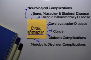
We knew since the mid 1990’s that chronic inflammation can cause many diseases. Among these are cancer, hardening of the arteries (atherosclerosis), arthritis, dementia (Alzheimer’s disease), frailty and other degenerative disorders. LifeExtension reviewed this topic in 2019.

Older people often accumulate chronic inflammation, which associates with the aging process. Persistent pain is one of the symptoms that chronic inflammation can cause.

Doctors use tests like the C-reactive protein, hemoglobin A1C, homocysteine and the erythrocyte sedimentation rate to measure chronic inflammation.

When the doctor diagnoses chronic inflammation, it is important to find ways to inhibit it and finally resolve it. Curcumin, ginger and non-steroidal anti-inflammatories are examples of measures that help inhibit inflammation. But it is only lately that scientists have found specialized pro-resolving mediators (SPM) that help to resolve chronic inflammation. They are polyunsaturated fatty acids, which health food stores offer as supplements.

Specialized pro-resolving mediators (SPM)

Researchers isolated several pro-resolving mediators that are metabolites of omega-3 fatty acids. This link discusses an experiment where researchers obtained synovial fluid from arthritis patients who took omega-3 fatty acids. They compared this to synovial fluid from volunteers who also took omega-3 fatty acid supplements. Within 4 weeks there was a big difference for the arthritis patients with higher levels of E- and D-series SPMs in synovial fluid and plasma. Dr. Silverman reviewed the action of SPMs here. He points out that they can be taken as supplements and that they are safe, because they do not have any side effects. Conditions such as asthma, irritable bowel syndrome and various musculoskeletal conditions with chronic inflammation respond very well to SPM supplements. Omega-3 fatty acid supplements (EPA) metabolize into the E-series resolvins.  On the other hand, Krill oil with Docosahexaenoic acid (DHA) metabolizes into protectins, maresins and the D-series resolvins.

Three vital tasks of pro-resolving mediators

SPMs fulfill three vital tasks, called remove, restore and renew.  First, they activate macrophages, which remove dead cells and cellular debris as a result of chronic inflammation. Second, they restore the healthy balance of inflammatory compounds and anti-inflammatory substances. Third, SPMs help renew tissues that were damaged by chronic inflammation.

Some examples where SPM’s were helpful resolving inflammation in health conditions

  • Alzheimer’s disease patients had reduced SPMs in cerebrospinal fluid of living patients and in the hippocampus of corpses who had Alzheimer’s disease.
  • A research group showed that defective SPM resolution can be responsible for missing resolution of atheromatous plaques in arteries. This leads to more and more hardening of arteries until a heart attack or stroke occurs.
  • With obesity or metabolic syndrome insulin resistance develops. Researchers found that certain SPM’s are missing in obesity, which causes chronic inflammation. However, they also found that precursors of SPMs such as 17-hydroxydocosahexaenoic acid (17-HDHA) can serve as a novel treatment agent to treat obesity-related complications.

Newer studies about SPMs

  • 62 patients with knee osteoarthritis had the precursor of resolvins, 17-HDHA tested in the blood. Compared to controls with no arthritis the 17-HDHA level in the arthritis patients was low. When levels of 17-HDHA were high, there was no pain in osteoarthritis patients.
  • Chronic periodontitis is a chronic inflammatory condition in the mouth. In a rabbit model this condition was cured with resolvin E1 derived from omega-3 fatty acids.
  • Delayed wound healing is typical for type 2 diabetes. In a mouse model with diabetic mice researchers inflicted skin wounds on mice. Subsequently they showed that with resolvins mice wounds healed much faster than in control mice.
  • Chronic kidney failure, liver impairment, diabetes, obesity and coronary heart disease showed abnormalities in the SPM system in humans. Potentially they will be healed with the help of resolvins or their precursors, which includes the omega-3 fatty acids EPA and DHA.
Chronic Inflammation Can Cause Many Diseases

Chronic Inflammation Can Cause Many Diseases

Conclusion

New compounds have been detected that derive from the omega-3 fatty acids EPA and DHA. They have the name specialized pro-resolving mediators (SPM). Included are the resolvins D1 and D2, the resolvins E1, E2 and E3 as well as the precursors 18-hydroxyeicosapentaenoic acid (18-HEPE) and 17-hydroxydocosahexaenoic acid (17-HDHA). The function of these compounds is to stimulate macrophages to clean up areas of chronic inflammation and remove cell debris and injured cells that cannot survive. While anti-inflammatories like ginger and curcumin only tone down the inflammation, SPMs help to resolve chronic inflammation. Various chronic clinical conditions were identified as being due to chronic inflammation. Chronic kidney failure, liver impairment, diabetes, obesity and coronary heart disease showed abnormalities in the SPM system. We may soon see alternative approaches treating these conditions with specialized pro-resolving mediators.

Jun
06
2020

Adequate Vitamin D Level Strengthens the Immune System

The Covid-19 coronavirus crisis is teaching us that an adequate vitamin D level strengthens the immune system.

When we age, our resistance to infections weakens, but this may be because our immune system needs more vitamin D3. I have reviewed the super powers of vitamin D3 before in 2014. In the past the thought was that the human body would need only 400 IU of vitamin D3 every day to cure rickets. And these were the daily vitamin D3 recommendations from medical authorities for several decades. Gradually it became known that for cancer prevention, infection prevention, cardiovascular illness prevention and for diabetes prevention much higher doses of vitamin D3 were necessary. As pointed out in the previous link, almost 50% of the world population is deficient in vitamin D. This is due to a lack of exposure to sunlight and due to inadequate supplementation with vitamin D3.

History of vitamin D

Dr. Adolf Windaus received the Nobel prize for chemistry in 1928. It was to acknowledge “… his studies on the constitution of the sterols and their connection with vitamins”. His work involved the metabolism of vitamin D and the precursors of vitamin D.

Rickets

As the above link shows, rachitic children were treated since the mid 1800’s with cod liver oil and since the early 1900’s also with ultraviolet light. But we know now that 400 IU of vitamin D3 per day is just enough to cure rachitic children, but it is not enough to strengthen the immune system to fight influenza viruses or the Covid-19 coronavirus. I will discuss further below what vitamin D blood levels are important to achieve a healthy state of the immune system.

Adequate vitamin D level strengthens the immune system

The immune system is very complicated and consists of many cell types that interact with each other and the rest of the body. It is important to recognize that the innate immune system immediately inactivates intruding viruses. But the vitamin D blood concentration has to be high enough. The acquired immunity consists of antibodies that are produced by B cells. The antibodies were produced during prior infections that you have survived and you are now immune to. However, other antibodies that circulate in your blood may have originated from vaccines you received in the past (whooping cough, measles, tetanus, diphtheria etc.). With the Covid-19 coronavirus it is the innate immunity that plays the biggest role until a vaccine will be found in the future.

Vitamin D is a hormone

This 2013 paper explains that vitamin D is a hormone that stimulates its own vitamin D receptor. This is a nuclear receptor that has close relations to the cell DNA and can stimulate more than 900 polypeptides. They are messenger molecules that are involved in a variety of physiological functions. One of the key functions is the immune system. This link explains that T cells that have vitamin D receptors can develop into cytotoxic T cells (also known as “killer T cells”). They are important in fighting cancer, but also parasites.

The key is that the hormone vitamin D can release more than 100 polypeptides that have the power to fight virus attacks including the Covid-19 coronavirus.

Three mechanisms how vitamin D works against the virus

The researchers outlined 3 mechanisms of how vitamin D works:

  • Maintaining tight epithelial junctions making it more difficult for the Covid-19 coronavirus to penetrate.
  • “Killing enveloped viruses through induction of cathelicidin and defensins.” These powerful antiviral polypeptides can kill viruses that have invaded the blood stream within 1 to 2 days.
  • “…And reducing production of proinflammatory cytokines by the innate immune system, thereby reducing the risk of a cytokine storm leading to pneumonia.” It is people who get the viral pneumonia that are at a high risk of death. By bringing the blood level up to the higher range of normal, between 50 and 80 ng/mL, patients that have encountered Covid-19 coronavirus are more likely to survive.

Two polypeptides, cathelicidin and defensins

Again, I like to emphasize that it is not vitamin D that has a direct effect on the virus. It is two polypeptides, cathelicidin and defensins, which are powerful antiviral polypeptides, that are released by vitamin D.

They can kill viruses that have invaded the blood stream and can eliminate the cytokine storm. This all happens very fast, within only 1 to 2 days. But you have to have an adequate vitamin blood level for this to occur (about 50-80 ng/mL).

Sources of vitamin D

First of all, vitamin D is readily absorbed from food. But there are not many foods that contain enough vitamin D for the immune system. The ones that contain vitamin D are as follows:

  • “Fatty fish, like tuna, mackerel, and salmon.
  • Foods fortified with vitamin D, like some dairy products, orange juice, soy milk, and cereals.
  • Beef liver.
  • Cheese
  • Egg yolks. “

Sun induced amount of vitamin D

Secondly, vitamin D can be synthesized in the skin from exposure to sunlight. But for this to happen all the necessary enzymes need to be present.  This link explains that many older people above the age of 65 have low vitamin D blood levels because of a lack of sun exposure and a lack of cutaneous synthesis because of enzyme issues.

Vitamin D supplements

The most reliable source of vitamin D are vitamin D3 supplements. When people supplement with the same dose of vitamin D3 there will be people who get higher vitamin D blood levels than others, as absorption in the gut is different for different people.  The ones who have relatively low vitamin D blood levels are often called “slow vitamin D absorbers”. But when the vitamin D3 dosage is increased even those people will reach the recommended high normal range (50-80 ng/mL).

Vitamin D blood level

The vitamin D blood test has the scientific name “25-hydroxy vitamin D level”. This is now the recognized gold standard for determining who is deficient or has normal levels with respect to vitamin D. The following 2013 publication has studied the vitamin D level of 1,470 healthy Swiss men and women, 60 years or older. Vitamin D levels were classified as severely deficient when the level was below 10 ng/mL. The vitamin D level was deficient between 10 and 20 ng/mL. The level was insufficient when between 21 and 29 ng/mL. A level above 30 ng/mL is normal.

8 % of the subjects were severely insufficient and 66% had insufficient vitamin D levels. Only 26.1% of the subjects had normal levels. Over 50% of healthy older Swiss (above the age of 70) had insufficient vitamin D levels.

Which vitamin D level is safe and which is not?

A peer-reviewed publication of the effects of vitamin D in health and disease contains 269 references.

What vitamin D level is optimal? This question was reviewed in this paper.

  • Below 15 ng/mL the immune system is paralyzed
  • With a level above 30 ng/mL the immune system is working
  • A level of 50-80 ng/mL has the immune system working optimally
  • Above 150 ng/mL toxic vitamin D levels start
  • With 300 ng/mL severe toxicity begins

Vitamin D toxicity

It is only with high levels of vitamin D (more than 150 ng/mL) that you have to worry about high calcium levels in the blood or kidney stones (toxic levels). But the key is to not exceed 80 ng/mL regarding the vitamin D blood level. This gives you a lot of flexibility before you reach toxic levels (above 150 ng/mL). For those who want more information, here is a thorough, peer reviewed publication about vitamin D toxicity with 59 references.

Vitamin D supplement compliance

The question is why not more people take adequate vitamin D3 supplements.  We know that vitamin D can prevent so many chronic diseases including serious viral infections. The answer is complex, but it includes a fear of the population of vitamin toxicity (kidney stone and high calcium levels). However, as pointed out before, this occurs only above a vitamin D level of 150 ng/mL. With proper vitamin D blood level monitoring you never reach toxic levels of vitamin D.

Denial

Denial likely is another major factor. People feel that if they have a balanced diet, they would be protected from vitamin D insufficiency. As pointed out before this is a grave error to think as our food does not contain sufficient vitamin D to strengthen our immune system.

False security with low doses of vitamin D

Finally, there are people who think that low doses of vitamin D, like 1000 IU of vitamin D daily, would be enough. But it is not enough. This is why testing vitamin D blood levels is so important. It is a reality check. The blood level must be in the high normal range (50-80 ng/mL). At this level the immune system functions optimally.

Compliance issues

In this context there was an interesting study done by LifeExtension, a company that publishes monthly health magazines. In this study the company examined the vitamin D blood levels of LifeExtension members. They are the ones who should be knowledgeable in how important it is to have good, preventative vitamin D blood levels. The study showed that 38% of the vitamin D test results were less than 30 ng/mL. In addition, 69% of the vitamin D tests were less than 40 ng/mL. Finally, 85% of the vitamin D test results were less than 50 ng/mL. What this means is that LifeExtension members were non-compliant when it came to taking regular adequate vitamin D3 doses. This resulted in levels that were too low for the majority to protect them from the Covid-19 coronavirus.

Covid-19 coronavirus infections and vitamin D blood level

There is a tight relationship between vitamin D blood levels and the strength of the immune system. Essentially, coronavirus mortality measures who is vitamin D deficient. Without enough vitamin D on board the virus penetrates into the blood stream and penetrates the lining of the respiratory tract. Next the cytokine storm develops, which leads to viral pneumonia. Higher doses of vitamin D3 will mitigate the course of Covid-19 coronavirus.

Adequate Vitamin D Level Strengthens the Immune System

Adequate Vitamin D Level Strengthens the Immune System

Conclusion

The Covid-19 coronavirus pandemic has taught us how important an intact immune system is to survive the virus when you get it. We do know for some time how closely related a good vitamin D level is with the functioning of the immune system. I have reviewed here what a desirable vitamin D level is and how we can achieve this with oral vitamin D3 supplements. The goal is to achieve a vitamin D level in the upper range of normal (50-80 ng/mL). With a level like this the virus cannot penetrate the mucous membranes of the respiratory tract and even if it did, it cannot produce a cytokine storm in the blood that would lead to the deadly viral pneumonia or to blood clots. When the virus invades the bloodstream, vitamin D releases powerful antiviral polypeptides that can kill viruses within 1 to 2 days.

Literature

Here are some peer-reviewed publications on vitamin D:


May
16
2020

Cutting Out Bad Lifestyle Habits Increases Life Expectancy

A Jan. 8, 2020 study stated that cutting out bad lifestyle habits increases life expectancy. It was a publication in the British Medical Journal by the Harvard T.H. Chan School of Public Health. The senior author of the study was Frank Hu, chair of the Department of Nutrition at Harvard Chan School. The researchers looked at data collected from a large group of males and females that had been accumulated for 34 years. They found that a 50-year-old male or female who did not change their lifestyle habits had another 25.5 years to live for males and another 29 years to live for females.

Five bad lifestyles and life expectancy 

The 5 bad lifestyle habits were smoking, a body mass index above 25.0, excessive alcohol intake, less than 30 minutes of exercise per day and an unhealthy diet.

When people cut out all of those risky lifestyles, 50-year-old men and women had a life expectancy of 37.6 years for men and 43.1 years for women. This was an addition of 12 years of healthy life for men compared to controls who did not change their lifestyles. With regard to women there was an addition of 14 years of life compared to controls.

Diseases that kill prematurely

The research team wanted to know what diseases caused the most reduction in life expectancy when people did not change their bad lifestyle habits. Men and women who cut out all negative lifestyle habits were 82% less likely to die from cardiovascular disease. They also were 65% less likely to die from cancer. Bad lifestyles caused the two major diseases, cardiovascular disease (heart attack and strokes) and cancer, which increased overall mortality. The overall study time was about 30 years. The third major disease that can cost lives is diabetes. In addition, the research team identified chronic diseases as being another potential cause of people dying prematurely.

Additional life expectancy

When life expectancies were broken down according to diseases, the following was noticed.

For men:

When cancer free    When free of heart issues   Without diabetes

6 more years             9 more years                         10 more years

For women:

When cancer free    When free of heart issues   Without diabetes

8 more years            10 more years                       12 more years

Dr. Frank Hu, who chairs the department of nutrition at Harvard T.H. Chan School of Public Health said: ”We found that following a healthy lifestyle can substantially extend the years a person lives disease-free”. Those who do not shed their bad lifestyle habits will come down with one or more of the mentioned diseases and die prematurely. Others who cut out all their bad lifestyle choices live substantially longer.

Diseases caused by poor lifestyle habits

It is important to review the diseases that shorten life expectancy due to having poor lifestyle habits.

Cardiovascular disease

Smoking, lack of regular exercise and poor eating habits result in being overweight or developing obesity. All of these are risks with LDL cholesterol elevation and HDL cholesterol lowering that leads to heart attacks and strokes. Here is a study that shows how life is shortened after a heart attack.

It is clear from this how important it is to give up all of the poor lifestyle habits to avoid this from happening.

Cancer

90% of lung cancers are the result of cigarette smoking.

Heavy drinking can contribute and also lead to cancer of the liver, esophageal cancer, cancer of mouth and throat and cancer of the breasts in women. In addition, cancer of the colon and rectum are also caused by consuming too much alcohol in both sexes.

Diabetes

There are a variety of risk factors causing diabetes. Obesity, a lack of exercise, a bad diet with too much carbohydrates and the aging process are what contributes to the development of type 2 diabetes.

We see again that it is largely lifestyle issues that drive the onset of this disease. People who have developed diabetes need to control their blood sugar very closely to avoid complications of diabetes. This includes making healthier choices.

Otherwise complications of diabetes are diabetic nephropathy, blindness from macular degeneration of the cornea, heart attacks, stroke and diabetic neuropathy. In addition, vascular complications also include artery occlusions in the lower extremities with frequent foot or below knee amputations.

Chronic diseases

Often chronic diseases develop when there is generalized development of inflammation. COPD, chronic kidney disease and arthritis are examples of such conditions. In addition, Alzheimer’s disease, arthritis, asthma, Crohn’s disease, cystic fibrosis and diabetes belong into this category. All of these chronic diseases have in common that cytokines produce inflammation in the body. This keeps the chronic disease going and makes it more difficult to cure. When the person with a chronic disease makes poor lifestyle choices, the inflammation just becomes more chronic.

Smoking is one of the factors that makes chronic inflammation more chronic. Having a body mass index above 25.0 (being overweight) and above 30.0 (obesity) also creates more inflammation in the body. Excessive alcohol intake damages body cells and releases free radicals. These in turn cause inflammation and make the chronic disease more difficult to treat. An unhealthy diet tends to raise the bad LDL cholesterol, introduces pesticides and other chemicals into the system and adds to the chronic inflammation. Finally, a lack of exercise is not contributing to a healthy circulation and lowers the protective HDL cholesterol, paving the way for heart attacks and strokes.

Cutting Out Bad Lifestyle Habits Increases Life Expectancy

Cutting Out Bad Lifestyle Habits Increases Life Expectancy

Conclusion

Bad lifestyle habits are what causes us to get illnesses and die prematurely. Functional medicine and anti-aging medicine are at the frontier of modern medicine. These specialties are teaching us how to stay well and age gracefully. For decades conventional medicine has treated only symptoms, but not the causes of diseases. But this has not changed the mortality rates of heart attacks and cancer. Smoking, a body mass index above 25.0, excessive alcohol intake, exercising less than 30 minutes of exercise per day and an unhealthy diet are causes that make us sick. These bad lifestyle factors cause chronic inflammation in our system. They are the real cause of heart attacks, strokes, cancer and diabetes, the major killer diseases that prevent healthy aging.

Facing bad lifestyle habits

Bad lifestyle habits cause chronic inflammation. T he C-reactive protein blood test can measure the degree of inflammation that is present. A fasting insulin level can detect whether a person is in danger of developing diabetes and improved lifestyle habits can prevent this from happening. Men can add 12 years and women 14 years of healthy life by eliminating bad lifestyle habits. By eliminating chronic inflammation our new life expectancy, which is 79 to 80 years, now can reach 92 to 94 years. It is up to you how healthy you stay and whether or not you age gracefully.

Incoming search terms:

Apr
25
2020

Exosomes can Regenerate Your Stem Cells

Dr. Douglas J. Spiel gave a talk on how exosomes can regenerate your stem cells. In essence, this was at the 27th Annual World Congress on Anti-Aging Medicine in Las Vegas from Dec. 13 to 15th, 2019. His original topic was: “Placental MSC Exosomes for Longevity and Chronic Disease”. Notably, MSC stands for “mesenchymal stem cells”. Dr. Spiel recommended this website to look at applications of exosome therapy.

Essentially, what scientist found is that certain factors from stem cells can activate your own stem cells to regenerate tissues that grow old. These factors are messenger RNA (mRNA) and micro RNA (miRNA), which come as tiny particles of 40‐100 nm.

Advantages of administering exosomes

To emphasize, exosomes can be given systemically as infusion, and they can regenerate your stem cells, if they are in need of treatment. They cross the blood brain barrier, so it is possible to treat brain diseases. That is to say, there is no first-pass removal in the lungs as it is with mesenchymal stem cells (MSC). The potency is related to the age of the donor and his/her stem cells. Notably, exosomes are easy to store, freeze and administer.

Exosomes influence the growth of target cells and promote regeneration. In addition, exosomes stimulate immunomodulation and have anti-inflammatory and anti-fibrotic properties. To clarify, the only limitations are that the strength of the exosomes is related to the age of the blood donor. The exosome fraction comes from mesenchymal stem cells. That is to say, it circulates in the plasma portion of the blood, which is obtained by spinning blood cells down in a centrifuge. To emphasize, exosomes can regenerate your stem cells.

Applications of exosomes for various clinical conditions

Joint inflammation

Mesenchymal stem cells are useful to treat arthritis. But it is important to realize that exosomes from mesenchymal stem cells are doing the same by stimulating the body’s own stem cells situated in the joints. In fact, several target cells have been identified that are stimulated by exosomes. These are chondrocytes, chondrocyte progenitor cells, cartilage-derived stem cells and synovium‐resident multipotent progenitor cells. In addition, other target cells are osteoblasts and osteoclasts in resident MSC within the subchondral bone and chondrogenic cells in the knee joint.

Disc degeneration  

Degenerative intervertebral discs respond to exosome treatments. The IL1 beta cytokine is involved in intervertebral disc degeneration. Exosomes inactivate these cytokines and have antioxidant and anti-inflammatory effects. Exosomes are not all the same. Different sub-fractions were isolated that have anti-inflammatory, immune-stimulating, antioxidant and other effects on the body.

Aging research

Researchers were able to pinpoint aging to various factors that contribute to premature aging. To clarify, when there is a decrease of catabolic processes and an increase of anabolic processes, an older person can combat premature senescence. Another key point, aging is also linked to redox homeostasis. Simply put, oxygenation processes in the body need to be balanced by reduction processes. This keeps the body in a healthy state. ADP/NADH production can be stimulated by exosomes.

Longevity comes from good lifestyles

With the use of exosomes, the aging process slows down, as oxidative stress is neutralized, damaged mitochondria are removed and cellular debris as well. That is to say, this improves inflammation and premature aging.

As has been noted, in the past 200 years life expectancy has doubled in most countries. 4 areas where longevity is particularly common are: Okinawa, Japan; Sardinia, Italy; Nicoya, Costa Rica and Loma Linda, USA. Only 7% of longevity stems from genetic factors, the rest is from lifestyles we adopt. In the final analysis, people who die prematurely followed a very poor lifestyle causing them to develop diseases, which ultimately killed them.

Clinical diseases from aging

Ultimately, advanced aging puts you at risk of getting cardiovascular disease (heart attacks and strokes), cancer and neurodegenerative diseases (Alzheimer’s disease, Parkinson’s disease). From the third decade onwards, there is the risk of bone loss, which causes osteoporosis. As has been noted, loss of cartilage causes osteoarthritis. Loss of muscle strength and muscle mass is called sarcopenia. With aging there is often an accumulation of abdominal fat. Hormones are disbalanced. Blood pressure is often elevated and blood lipids as well. Insulin resistance can develop and the blood vessels become stiffer. This causes heart attacks and strokes.

The details of the aging process are much more complicated than originally thought of. There is a combination of aging of the DNA, mitochondrial aging, stem cell exhaustion and a change of intercellular communication due to dysregulated endocrine signalling. In addition, there is a decline of the immune system and epigenetic factors that can turn off longevity genes.

Oxidative stress as a cause of premature aging

Dr. Spiel pointed out that reactive oxidative species (also known as free radicals) cause damage to mitochondria and mitochondrial DNA. But we need the energy from the mitochondria for a comfortable life. In essence, antioxidants can neutralize free radicals. Age-related conditions due to oxidative stress are: cardiovascular disease, chronic kidney disease and type 2 diabetes, chronic obstructive pulmonary disease, cancer, neurodegenerative disease, frailty and sarcopenia. Surely, both reactive oxygen and reactive nitrogen are free radicals. They have one or more unpaired electrons and all aerobic body cells produce them. Reactive oxygen and nitrogen species (RONS) cause oxidative damage to our cells and contribute to the development the diseases just mentioned.

Antioxidants help to prevent diseases

But antioxidants can contain these free radicals in various ways. The body has five built-in enzymatic ways to protect itself and five non-enzymatic ways (bilirubin, vitamin E, beta-carotene, albumin and uric acid). In addition, there are antioxidants that a person can take as supplements to inactivate RONS. These are: vitamin C and E; phenolic antioxidants like resveratrol, phenolic acids, flavonoids, oil lecithin, selenium, zinc and drugs like acetylcysteine.

Without control of the oxidative stress RONS can lead to cellular senescence and chronic inflammation. This leads to a vicious cycle where chronic oxidative stress and inflammation feed on each other leading to premature diseases.

Causation of several diseases

As we age, the body reduces the inborn antioxidant enzymes (superoxide dismutase and glutathione peroxidase). Before we can understand how to live longer, we need to be aware what happens in various health scenarios as follows.

  • The lack of inborn antioxidant enzymes leads to vascular endothelial dysfunction, high blood pressure and premature hardening of the arteries. This can become a precursor to heart attacks and strokes.
  • Elevated blood sugar in the case of type 2 diabetes leads to increased sugar concentration of body cells and formation of free radicals.
  • Oxidants from cigarette smoke activate macrophages and epithelial cells to produce inflammatory cytokines. Continued smoking releases proteases in the process that break down connective tissue and cause emphysema and COPD.

There are more diseases

  • Chronic kidney disease comes from oxidative stress affecting the filter units of the kidney, called glomeruli. With a lack of blood supply to the kidneys secondary high blood pressure develops and endothelial dysfunction. It also leads to chronic inflammation.
  • In the brain oxidative stress leads to cognitive impairment and dementia.
  • Oxidative stress and chronic inflammation are important ingredients for the development of cancer. RONS and cytokines release NF-kB, which activates cancer genes. RONS can also directly attack the DNA of cells and cause cancer through carcinogenesis.
  • Sarcopenia and frailty come from the action of RONS on the skeletal muscles. In old age there are less inborn antioxidants available. This leads to decreased muscle quantity or sarcopenia. Eventually frailty results with the risk of falls and fractures. 

Preventative measures for slowing the aging process

There is a number of steps that in combination help to slow the aging process.

  • A Mediterranean diet combined with a fasting mimicking diet or other calorie restricted diet
  • Regular physical activity
  • Cognitive training
  • Vitamin D3 supplementation
  • Reducing your risk to develop vascular disease
  • Certain drugs turn on the longevity gene (metformin, rifampin)
  • Spiel warned that due to limited compliance and variable response these steps alone may not be enough to prevent age-related problems

How to live longer

It is important to recognize the importance of antioxidants to counteract the development of these diseases. As already mentioned, the following counter the effect of free radicals: vitamin C and E; phenolic antioxidants like resveratrol, phenolic acids, flavonoids, oil lecithin, selenium, zinc and drugs like acetylcysteine. Mesenchymal stem cells can also stop the action of free radicals. In addition, exosomes, which  are products of mesenchymal stem cells can do the same. Mitochondria, the power houses within the cells, create energy, but also release free radicals. In his clinic Dr. Spiel administers intravenous exosomes to counter the oxidative stress. Numerous studies linked mitochondrial dysfunction to various age-related diseases. There are markers in blood tests that the physician can order to analyze malfunctions in the body. Dr. Spiel showed 4 slides that contained a lot of medical information that is too technical. I omitted it for this review.

Intravenous infusions of exosomes

The important thing to remember is that epigenetics can be changed by exosome infusion and lifestyle changes mentioned above. Dr. Spiel said that generally he uses 15 ml of exosomes by intravenous infusion every 12 weeks for longevity and performance enhancement. This treats conditions like infertility, osteoporosis, osteopenia, heart, liver and kidney weaknesses. Here is the dosing for intravenous exosomes by weight:

20-50 lb: 5 ml; 50-90 lb: 10ml; more than 90 lb: 15 ml; more than 220 lb: 20 ml. Unfortunately, one exosome treatment costs between 500.00 and 922.00 USD, an amount that most people cannot afford.

Contraindication to the use of stem cells or exosome therapy

It is important to realize that a person who has cancer should not receive either mesenchymal stem cells or exosomes. Indeed, exosomes do not differentiate between cancer cells and healthy cells, but stimulate cell division. For the same reason people with myeloproliferative disease (sickle cell anemia, bone marrow dysplasia) should also not receive exosomes. To clarify, other conditions where the physician will not order exosomes are primary pulmonary hypertension, acute bacterial infection or an immune-compromised state. In addition, macular degeneration with neovascularization is also a condition where the health professional does not administer exosomes.

Exosomes can Regenerate Your Stem Cells

Exosomes can Regenerate Your Stem Cells

Conclusion

Dr. Douglas J. Spiel gave a talk on how exosomes can regenerate your stem cells. Specifically, this was at the 27th Annual World Congress on Anti-Aging Medicine in Las Vegas from Dec. 13 to 15th, 2019. Dr. Spiel explained how disease processes age our organs. Reactive oxygen and nitrogen species (RONS) cause oxidative damage to our cells and contribute to the development of diseases. This involves the mitochondria in the cells as well. The good news is that a healthy lifestyle can counter these damaging processes to a certain extent. But it takes another step to re-establish the balance of our cells, exosome infusions. Exosomes are tiny particles that are shed by stem cells and that circulate in the blood. They can reenergize stem cells that are ailing to become functional again.

Expensive exosome infusions

He recommended an infusion with exosomes every 12 weeks for maintenance of good health and as a “fountain of youth”. Obviously, there are some limitations. As mentioned, it is not suitable for all patients, like cancer patients, patients with sickle cell anemia, acute bacterial infections or pulmonary hypertension. In addition, it is also not a treatment which many patients will seek out as the cost is prohibitive. One exosome treatment cost between 500.00 and 922.00 USD, an amount that most people cannot afford.

Apr
18
2020

Changes of Metabolism by Inflammation

Dr. James LaValle gave a presentation about changes of metabolism by inflammation in Las Vegas. I listened to this lecture on Dec. 15, 2020. The 27th Annual World Congress on Anti-Aging Medicine in Las Vegas took place from Dec. 13 to 15th, 2019. His original title was: “Innovations in Metabolism and Metaflammation”. This talk was complex and as a result it may not be easy reading. But it shows how various factors can affect our metabolism and our life expectancy.

In the first place he understands “metabolism” as all of the chemical reactions together that make you feel the way you feel today. In the same way metabolism is the chemistry that drives you toward future health. It is equally important to note that disregulation of your metabolism occurs from global metabolic inflammatory signalling. As has been noted he called this “metaflammation” (inflammation affecting your metabolism).

Dr. LaValle said that understanding disruptors of your metabolism can lead to renew your health on a cellular level. The key to achieve this is to remove inflammatory signals.

Factors that accelerate aging and damage your metabolism

It is important to realize that several factors interfere with the normal aging process. Oxidative stress and inflammation are major factors. But hormone disbalance and increased blood sugar values and insulin resistance can also contribute to accelerated aging and damage your metabolism. Certainly, with a disturbance of the immune balance, autoimmune reactions can take place, which also does not help. In addition, pollutants from the environment derange the metabolism due to heavy metals that block important enzymatic reactions. In the minority there are also genetic factors that can interfere with a normal metabolism.

Many of the metabolic changes can lead to chronic inflammation. One source of inflammation can be lipopolysaccharides that stimulate the immune system to start an inflammatory process.

Many conditions are associated with inflammation such as diabetes, obesity, stress, the SAD diet (standard American diet), and liver or kidney damage.

How Metaflammation is developing

Metaflammation can start in the gut with microbiota alterations. The wrong types of bacteria can release lipopolysaccharides, and low grade endotoxemia develops. With obesity inflammatory kinins start circulating in the body. Stress can activate inflammatory substances in the brain and the rest of the body. Major contributors to inflammation in the body come from faulty diets. The Western diet contains too much sugar and refined carbs; it is too high in trans fats and saturated fats. It contains too many artificial additives, preservatives, salt, sweeteners and dyes. And it is too low in nutrients, complex carbs and fiber.

More problems with metaflammation

Kidney and liver illness can contribute to metaflammation. Several diseases come from chronic inflammation, like cardiovascular disease, type 2 diabetes, chronic kidney disease, depression, cancer, dementia, osteoporosis and anemia. Metaflammation alters the methylation patterns, which can slow down your metabolism. Increased blood lipids and chronic inflammation of the blood vessels lead to cardiovascular problems. The liver and kidneys are the major detoxification organs, and their disease leads to more metaflammation. Metaflammation also leads to hormone disbalances, sleep disorders and dysfunction of the immune system. The brain reacts to metaflammation with cognitive dysfunction and mood disorders. Muscle loss (sarcopenia) is another issue, so is osteoporosis. Finally, chronic metaflammation can cause cancer.

Major causes of metaflammation

The three major causes of metaflammation are changes of the gut microbiome, obesity and chronic stress. When the gut bacteria change because of a Western diet, the wrong bacteria release lipopolysaccharides that are absorbed into the blood. The gut barrier is breaking down and a low grade endotoxemia develops. With obesity adipokines, which are inflammatory substances secreted by the fatty tissue, circulate in the blood. Chronic stress activates inflammation in the brain and in the body.

Two major conditions are common with metaflammation: hyperlipidemia (high fat levels in the blood) and hyperglycemia. Both of these conditions change the metabolism and lead to cardiovascular disease (hyperlipidemia) or to type 2 diabetes (hyperglycemia). Both of these metabolic changes lead to one or more of the conditions mentioned above, accelerate the aging process and lead to premature deaths.

Interaction of various organ systems can cause metaflammation

Dr. LaValle stated that it is vital that your hormones stay balanced. With chronic stress cortisol production is high. This causes increased insulin production, reduced thyroid hormone and lowered serotonin and melatonin production in the brain. It also leads to autoimmune antibodies from the immune system and decreased DHEA production in the adrenal glands. In addition, growth hormone production and gonadotropin hormones are slowing down. We already heard that cortisol levels are up. The end result of these hormone changes is that the blood pressure is up and abdominal visceral obesity develops. The brain shows cognitive decline, with memory loss as a result. The bones show osteopenia, osteoporosis and fractures. The muscles shrink due to sarcopenia, frailty is very common. Heart attacks and strokes will develop after many years. The immune system is weak and infections may flare up rapidly. There are also higher death rates with flus.

Other mechanism for pathological changes with hormone disbalances

When Insulin is elevated, inflammatory markers are found in the bloodstream. This elevates the C-reactive protein and leads to damage of the lining of the blood vessels in the body. A combination of insulin resistance and enhanced atherosclerosis increases the danger for heart attacks or strokes significantly.

There is a triangle interaction between the thyroid, the pancreas and the adrenals. Normally the following occurs with normal function. The thyroid increases the metabolism, protein synthesis and the activity of the central nervous system. The pancreas through insulin converts glucose to glycogen in the liver. It also facilitates glucose uptake by body cells. The adrenal hormones are anti-inflammatory, regulate protein, carbohydrate and lipid metabolism and contribute to energy production.

Change of thyroid/pancreas/adrenals triangle when cortisol is elevated

When cortisol is elevated the balance of the thyroid/pancreas/adrenals’ triangle is severely disturbed. Cortisol is high, the T4 to T3 conversion is limited and, in the brain, there is hippocampus atrophy with memory loss and brain fog. The immune system will change with production of inflammatory kinins (IL-6 and TNF alpha). Insulin sensitivity is down, sugar craving up and weight gain develops (central obesity).

Change of thyroid/pancreas/adrenals triangle when the thyroid is depressed

The thyroid activity can be lower because of autoimmune antibodies (Hashimoto’s disease) or because of hypothyroidism developing in older age. This leads to decreased pregnenolone synthesis from cholesterol. As pregnenolone is the precursor for all the steroid hormones, the metabolism slows down profoundly. Mentally there is depressed cognition, memory and mood. The cardiovascular system shows reduced function. In the gut there is reduced gastric motility. The mitochondria, which are tiny energy packages in each cell, are reduced in number, which causes a loss of energy. There is increased oxidative stress, increased lactic acid production and decreased insulin sensitivity.

Cardiovascular disease not just a matter of high cholesterol

Dr. LaValle stressed that a heart attack or stroke is not just a matter of elevated cholesterol. Instead we are looking at a complicated interaction between hypothyroidism, diabetic constellation and inflammatory gut condition. The inflammatory leaky gut syndrome causes autoimmune macrophages and Hashimoto’s disease. The end result is hypothyroidism. The inflammatory kinins (TNF-alpha, IL-6) affect the lining of the blood vessels, which facilitates the development of strokes and heart attacks. You see from this that cardiovascular disease development is a multifactorial process.

Microbiome disruption from drugs

Drugs affecting the intestinal flora are antibiotics, corticosteroids, opioids, antipsychotics, statins, acid suppressing drugs like protein pump inhibitors (PPI’s) and H2-blockers. Other factors are: high sugar intake, pesticides in food, bactericidal chemicals in drinking water, metformin, heavy metals and alcohol overconsumption. Chronic stomach infection with H. pylori, stress and allergies can also interfere with the gut microbiome.

The microbiome disruption affects all facets of metabolism. This means that there can be inhibition of nutrient absorption and this may affect the gut/immune/brain axis. There are negative effects on blood glucose levels and insulin resistance. A disturbance of the sleep pattern may be present. A significant effect on the hormonal balance can occur (thyroid hormones, sex hormones and appetite related hormones). When liver and kidney functions slow down, there is interference of body detoxification.

Dr. LaValle talked more about details regarding the gut-brain-immune pathology. I will not comment on this any further.

Changes of Metabolism by Inflammation

Changes of Metabolism by Inflammation

Conclusion

Dr. LaValle gave an overview in a lecture regarding changes of metabolism by inflammation. This took place at the 27th Annual World Congress on Anti-Aging Medicine in Las Vegas from Dec. 13 to 15th, 2019.

This article is complex and contains a lot of detail, but there is one simple truth: oxidative stress and inflammation are major factors that influence our health on many parameters and lead to a list of illnesses. They lead to hormone disbalance and increased blood sugars and insulin resistance, which can also contribute to accelerated aging and damage of your metabolism. Dr. LaValle explained how high cortisol from chronic stress can lead to low thyroid hormones and in the brain, there is hippocampus atrophy with memory loss and brain fog. With alterations of the immune system there is production of inflammatory kinins (IL-6 and TNF alpha). Insulin sensitivity is down, sugar craving up and weight gain develops (central obesity). It does not stop there! We put our hope in medications, but the sad truth is that there are

Drugs that change the gut biome

Many drugs that are common also change the gut biome with resulting increased permeability of the gut wall (leaky gut syndrome). This overstimulates the immune system and leads to autoimmune diseases like Crohn’s disease and rheumatoid arthritis. Whenever there is an injury to the gut barrier, the blood brain barrier is following suit. This is how brain disease can develop as a result of a change in the gut biome. Impaired cognition, memory and mood can result from this. Alzheimer’s disease is one of the worst conditions that may be related to a combination of gut inflammation, chronic stress and inflammatory kinins.